
The parotid gland, and what a parotidectomy is
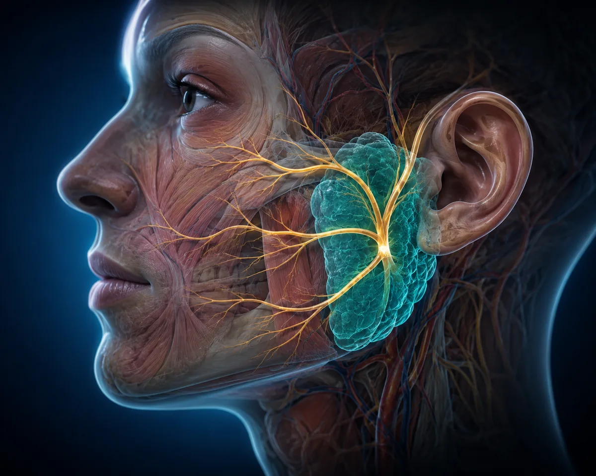
You have two parotid glands — the largest of the salivary glands — sitting just in front of and below each ear, wrapped around the angle of the jaw. They produce much of the saliva you swallow without ever thinking about it.
A parotidectomy is the operation to remove part or all of that gland, almost always because a lump has developed inside it. The reason this particular operation demands an experienced surgeon is not the gland itself. It is what runs straight through it: the facial nerve, the nerve that controls every muscle you use to smile, blink and raise an eyebrow.
The facial nerve enters the parotid and fans out through it like the branches of a tree, splitting the gland into a superficial and a deep part. A tumour cannot simply be scooped out. The surgeon must first identify the nerve trunk, then carefully trace each branch and dissect the gland away from it. That dissection is the operation.
Symptoms you should not ignore
Parotid tumours are typically undramatic, which is exactly why they get left. See an ENT and head & neck surgeon if you notice:
The first three are the common presentation and usually indicate a benign tumour. The remaining features are the ones that raise concern about a malignant tumour and warrant prompt assessment rather than a wait-and-watch approach.
Facial weakness alongside a parotid lump is always a reason to be seen quickly. A benign tumour pushes the facial nerve aside; it does not usually stop it working.
Why parotid lumps need a specialist opinion
Roughly eight in ten parotid tumours are benign, most commonly a pleomorphic adenoma. That statistic is reassuring, and it is also the reason these lumps get neglected for years. Three things are worth understanding:
- Benign does not mean harmless. A pleomorphic adenoma left alone continues to grow, becomes technically harder to remove cleanly, and over many years carries a small but genuine risk of malignant change.
- An incomplete first operation is difficult to correct. If a tumour is shelled out rather than removed with an adequate cuff of surrounding gland, it tends to recur — often in multiple nodules scattered through scar tissue, with the facial nerve now embedded in that scar. Revision surgery is markedly more hazardous than a correct first operation.
- Not every lump in that area is a parotid tumour. Lymph nodes, cysts, infections and skin lesions all present similarly and are managed completely differently.
What happens before any surgery is discussed
No responsible surgeon proposes a parotidectomy on examination alone. The workup usually includes:
- Clinical examination. The lump is assessed for size, mobility, consistency and skin involvement, along with a careful test of facial nerve function and an examination of the neck for enlarged nodes.
- Ultrasound. First-line imaging — quick, no radiation, and good at distinguishing solid tumours from cysts and lymph nodes.
- FNAC. Fine needle aspiration cytology, where a thin needle takes a sample of cells for the pathologist. It is done in the clinic with local anaesthetic and is the key test for distinguishing benign from malignant before planning.
- MRI or CT. Used for larger tumours, anything suspected of involving the deep lobe, or where FNAC raises concern — it maps the tumour's exact relationship to the nerve and deeper structures.
Only once those results are in can a realistic conversation happen about whether surgery is needed, which operation, and what the likely outcome is.
Types of parotidectomy
| Operation | What is removed | Typically used for |
|---|---|---|
| Superficial parotidectomy | The part of the gland lying superficial to the facial nerve | The majority of benign tumours, which sit in the superficial lobe |
| Partial / extracapsular dissection | The tumour with a cuff of surrounding normal gland | Small, well-defined, clearly benign tumours in selected patients |
| Total parotidectomy | Both superficial and deep lobes, facial nerve preserved | Deep lobe tumours, and many malignant tumours |
| Radical parotidectomy | Gland together with the facial nerve, with nerve grafting considered | Uncommon — advanced cancers already invading the nerve |
Superficial parotidectomy is by a clear margin the most frequently performed of these, because that is where most tumours sit. Where a neck node is involved, a neck dissection may be performed at the same time — see head and neck surgery.
The facial nerve — the question everyone asks first
Almost every patient asks the same thing: will my face be affected? It deserves a straight answer.
In a planned operation for a benign tumour, the aim is always to preserve the facial nerve completely, and in experienced hands that is achieved in the large majority of cases. What is common — and should be expected rather than feared — is temporary weakness. Handling and stretching the nerve during dissection can leave it bruised, producing a weaker smile or an eyelid that does not close fully. This typically recovers over weeks to a few months.
Permanent weakness is uncommon in benign disease. The risk rises with revision surgery, large deep-lobe tumours, and malignant tumours that have grown into the nerve. Where cancer has directly invaded the nerve, removing it may be unavoidable — and in that situation nerve grafting and reanimation options are discussed openly beforehand.
During surgery a facial nerve monitor is used, which alerts the surgeon when instruments come close to nerve fibres. It does not replace careful dissection, but it is a valuable additional safeguard.
What the operation involves
Parotid surgery is performed under general anaesthesia and usually takes somewhere between two and four hours, depending on tumour size and position.
- The incision. A modified Blair incision runs in the natural crease in front of the ear, curves under the earlobe and into the upper neck — placed to follow existing skin creases and hairline so it settles into an inconspicuous line.
- Identifying the nerve. The main facial nerve trunk is located at a defined anatomical landmark, then each branch is traced forward under magnification.
- Removing the tumour. Gland tissue is dissected off the nerve branches and removed with an appropriate margin, keeping the tumour capsule intact.
- Closing. A small suction drain is usually placed for a day or two and the skin closed in layers.
Most patients stay one night in hospital. Complex surgery for malignancy may require longer. Major surgery is carried out at Fortis Hospital, Manesar, where Dr. Vardhan holds an association.
Recovery, week by week
- First 48 hours. Some swelling and discomfort, controlled with ordinary pain relief. The drain is removed once output settles, usually the next day.
- Week 1. Home and largely self-caring. Numbness of the earlobe and the skin over the operated area is normal and expected. Stitches are removed around day 7.
- Weeks 2–4. Most people return to desk-based work within two weeks. The scar is pink and firm at this stage — this is normal healing.
- Months 2–6. Any temporary facial weakness continues improving. The scar softens and fades steadily.
- Beyond 6 months. The scar continues to mature for up to a year. Earlobe numbness may persist to some degree long-term, and most patients find it a minor issue.
Follow-up is important. Histology from the removed specimen is reviewed with you, and where the tumour was malignant, a surveillance schedule is arranged — occasionally alongside oncology input for radiotherapy.
Risks, stated honestly
Every operation carries risk, and you are entitled to hear these before consenting rather than afterwards:
- Temporary facial weakness — relatively common, usually resolving over weeks to months.
- Permanent facial weakness — uncommon in benign disease; risk is higher in revision, deep lobe and malignant cases.
- Numbness of the earlobe — very common, from division of the greater auricular nerve. Often partially recovers.
- Frey's syndrome — sweating or flushing of the cheek when eating, appearing months later as nerve fibres regrow along altered pathways. Usually mild; treatable if troublesome.
- Salivary fistula or sialocele — a collection or leak of saliva at the wound, generally settling with simple measures.
- Haematoma or infection — uncommon, managed in the usual way.
- Recurrence — low after an adequate first operation; considerably higher after incomplete removal, which is precisely why the first operation matters so much.
Choosing a surgeon for parotid surgery
This is not an operation to select on price or convenience. Reasonable questions to ask any surgeon:
- How many parotidectomies do you perform, and are you a trained head and neck surgeon?
- Will FNAC and imaging be done before we decide on surgery?
- Do you use facial nerve monitoring during the procedure?
- What is your plan if the histology comes back malignant?
- Where will the incision be placed, and who follows me up afterwards?
Dr. Harsh Vardhan is an ENT specialist and head & neck surgeon with over 2,000 operations performed, whose declared areas of interest include head and neck tumour surgery and endoscopic skull-base work. He is a member of the Foundation of Head and Neck Oncology and Joint Secretary of the AOI Gurugram Branch. Assessment, FNAC and follow-up take place at the Sector 82A clinic; surgery is performed at Fortis Hospital, Manesar.
Patients travel to the clinic from across New Gurugram, Kherki Daula, Manesar, Badshahpur, Sohna Road and the Dwarka Expressway corridor.